A 31-YEAR-OLD FEMALE SUFFERING FROM RARE UNILATERAL TEMPOROMANDIBULAR JOINT ANKYLOSIS GETS A NEW LEASE OF LIFE WITH ADVANCED 3D IMPLANT AT S. L. RAHEJA HOSPITAL MAHIM
For the first time in Mumbai, the Temporomandibular Joint (TMJ) replacement surgery was performed using a 3D replica and customized implant ~ Saba Shaikh, aged 31, faced significant difficulty in opening her mouth and chewing food

For the first time in Mumbai, the Temporomandibular Joint (TMJ) replacement surgery was performed using a 3D replica and customized implant ~
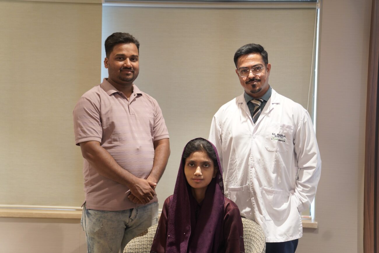
Saba Shaikh, aged 31, faced significant difficulty in opening her mouth and chewing food as a result of stiffness in her temporomandibular joint (TMJ). For more than a year, the patient depended on a liquid diet and utilized a flat spoon for eating. This gradually affected both her physical and mental well-being, as well as impeding her ability to speak clearly. A team of experts led by Dr Tofiq Bohra, Consultant – Oral & Maxillofacial Surgery Department at S. L. Raheja Hospital Mahim – A Fortis Associate implemented an innovative strategy involving Virtual Surgical Planning (VSP) for replacing the jaw joint affected by Bony Ankylosis. A 3D replica of the patient’s skull was created, that helped the surgeon in visualizing the expected results of the surgical procedure.
Saba, resident of Navi Mumbai was diagnosed with Nasopharyngeal Cancer four years ago, and had made successful recovery through radiation & chemotherapy. Subsequently, she started experiencing pain in her TMJ and encountered difficulty in opening her mouth. TMJ plays a crucial role in daily activities such as opening & closing the mouth and chewing food. She consulted multiple experts and also underwent an Open Joint Surgery of TMJ to address the issue. Unfortunately, this procedure proved unsuccessful. and resulted in Ankylosis (abnormal stiffening) of the TMJ.
As time passed, her ability to open her mouth gradually diminished, leading to complete lockjaw. She began experiencing substantial weight loss; she also experienced dental problems (like tooth decay and gum swelling) as she was unable to uphold oral hygiene.
She sought medical assistance at S. L. Raheja Hospital, Mumbai, when her mouth opening had further decreased, to less than the width of one finger, with only the right joint functioning and the left joint completely ankylosed (becoming stiff). Dr Tofiq Bohra examined her and then ordered CT and MRI scans of both sides of her jaw joint. The results showed that the left side was stiff and eroded, while the right side was worn down.
The patient’s CT scan and MRI findings were thoroughly studied, and a 3D replica of the patient’s skull was developed. The team of doctors suggested that the patient undergo Temporomandibular Joint (TMJ) replacement surgery; upon consent, the design of the implant was created on a 3D printer. On March 26, 2024 TMJ prosthesis was implanted. The prosthesis was then covered with a layer of the patient’s abdominal fat to provide extra padding above the joint, helping prevent damage to the prosthesis.
Dr Tofiq Bohra, Consultant – Oral & Maxillofacial Surgery Department at S. L. Raheja Hospital Mahim – A Fortis Associate said, “TMJ replacement surgery represents an innovative approach that surpasses alternative surgical methods by providing patients with a fresh prosthetic joint in the facial region, thereby minimizing the risk of Ankylosis recurrence. One hurdle encountered during this process was inserting a breathing tube for anesthesia due to limited mouth opening. However, this issue was effectively resolved through nasal endoscopy conducted with the aid of an anesthetist. The presence of fibrosis resulting from previous joint replacement surgery added another level of complexity to the surgery.
The patient’s husband, Azharuddin, said, “We were deeply concerned about my wife’s condition and her inability to open her mouth to eat or talk. If not treated on time, it could have led to other medical complications, making recovery even tougher. However, thanks to the team of doctors including Dr Ujwala Karmarkar and Dr Meghna Nadkarni, Saba is recovering well. She has started speaking near-normally, and we hope she’ll be able to enjoy her favourite food.”
Following the surgery, the patient showed promising progress within 48 hours and her mouth gradually opened up to two fingers width. She was prescribed a liquid diet for the first three days and later was shifted to a soft diet. Facial physiotherapy was started for further improvement without any facial nerve damage. Presently, the patient is faring well and can eat a soft diet with constant improvement in joint performance.


