IIT Jodhpur led multi-institutional team finds effective fluorescent probe for the diagnosis of Alzheimer’s disease
Ø The research was done by IIT Jodhpur in collaboration with IIT Kharagpur and CSIR-IICB, Kolkata Ø The diagnosis method of Alzheimer’s disease will provide an inexpensive reliable alternative Ø The developed probes enable rapid

Ø The research was done by IIT Jodhpur in collaboration with IIT Kharagpur and CSIR-IICB, Kolkata
Ø The diagnosis method of Alzheimer’s disease will provide an inexpensive reliable alternative
Ø The developed probes enable rapid and safe analytical sensing due to the absence of radioactive chemicals
A multi-institutional team led by Indian Institute of Technology Jodhpur has developed an efficient fluorescent molecular probe that can be used in the diagnosis of Alzheimer’s disease. The research has been carried out in collaboration with Indian Institute of Technology Kharagpur, and Council of Scientific & Industrial Research – Indian Institute of Chemical Biology, Kolkata.
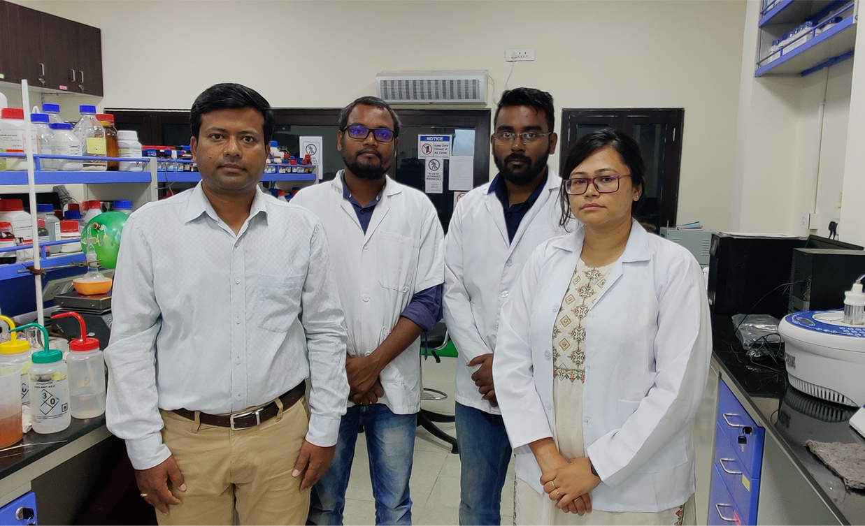
The research results have been recently published in ACS Chemical Neuroscience journal, in a paper co-authored by Dr. Surajit Ghosh, Professor, Department of Bioscience & Bioengineering, IIT Jodhpur along with his research scholars Mr. Rathnam Mallesh, Ms. Juhee Khan, and Mr. Rajsekhar Roy and Prof. Nihar Ranjan Jana, Head, Department of Biosciences, IIT Kharagpur, Dr. Parasuraman Jaisankar, Head, Organic & Medicinal Chemistry Division, CSIR – Indian Institute of Chemical Biology, Kolkata and Dr. Krishnangsu Pradhan, Senior Research Fellow, CSIR-Indian Institute of Chemical Biology, Kolkata.
According to the Dementia India Report 2010 by the Alzheimer’s and Related Disorders Society of India (ARDSI), there will be around 7.6 million Indians with Alzheimer’s and other dementia conditions by 2030 in India. Alzheimer’s disease is believed to be caused by the abnormal build-up of plaques in and around brain cells. Plaques are aggregates of a type of small protein (peptide) called amyloid-beta (Aβ).
The diagnosis of Alzheimer’s disease involves assessment of cognitive functions, the observation of brain size and structure through SPECT, PET and MRI scans, and the detection of amyloid plaques. Amyloid plaques are detected by extracting the brain fluid (cerebro-spinal fluid) through a spinal tap, or by PET Scans. Although both methods are reliable, they are invasive and expensive.

>Highlighting his research, Dr. Surajit Ghosh, Professor, Department of Bioscience & Bioengineering and Dean, Research and Development, IIT Jodhpur said, “Optical imaging systems that use fluorescent or colour-based chemicals to target tissues and molecules of interest are considered better diagnostic techniques in the biomedical area,” He added that fluorescence probes can enable rapid and safe analytical sensing due to the absence of radioactive chemicals or expensive equipment.
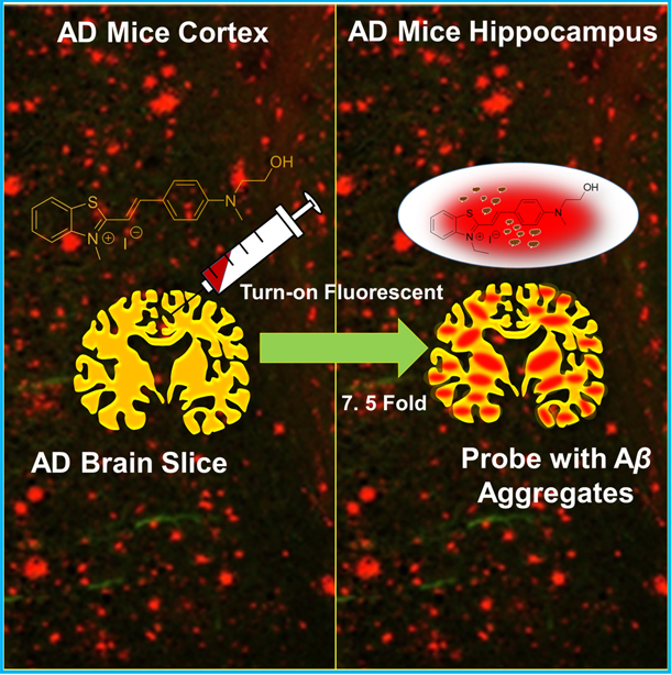
Fluorescent probe diagnosis involves injecting a fluorescent chemical that specifically attaches itself to the amyloid plaque and assessing the change in fluorescent properties using an appropriate detector. The fluorescent chemical, in addition to being able to specifically and selectively attach to Aβ aggregates, must also be able to cross the blood-brain barrier to reach the brain. There must also be a change in its fluorescent properties – colour and intensity – when it attaches to Aβ aggregates. Furthermore, the fluorescence must be in the visible light region for easy detection. The commercially available fluorescent probe ThT has poor blood-brain barrier crossing ability and exhibits only a 2.5-fold change in fluorescence intensity.
“We studied the binding modes of RM-28 with Aβ aggregates using molecular docking experiments and found that this probe binds to the entry site, inner clefts and surface of the Aβ aggregates. RM-28 can potentially replace ThT in the detection of Aβ aggregates in both in vitro and in vivo diagnostic techniques and could also serve as template molecular scaffold for designing novel or new fluorescent probes,” said Dr. Ghosh on the future implications of their discovery.
The researchers have successfully designed and developed a series of benzothiazole based fluorescent molecules that can selectively bind to Aβ aggregates. All these molecules were seen to emit fluorescence in one colour when unbound, and the emission colour shifted towards red in the visible light (rainbow – violet indigo blue green yellow orange red) spectrum with a concomitant increase in fluorescence intensity. For example, a molecule they named RM28 was yellow when unbound and orange when it was bonded to the Aβ aggregates, and there was a 7.5-fold increase in fluorescence intensity after binding. They used AD mice brain samples to test RM28.
This molecule was stable in biological fluids and could easily traverse the blood-brain barrier. It was also selective to Aβ aggregates in the presence of competing biomolecules. Hence, the probe found by the research team will provide a non-invasive and inexpensive reliable alternative to a spinal tap or PET Scan methods Alzheimer’s diagnosis.


