Transition from Analogue to Digital C-Arms
A Mobile C-Arm is sophisticated medical imaging equipment that finds widespread use in various operating room settings for image guided surgeries. C-Arms are based on X-ray technology and provide high-resolution X-ray images in real time
A Mobile C-Arm is sophisticated medical imaging equipment that finds widespread use in various operating room settings for image guided surgeries. C-Arms are based on X-ray technology and provide high-resolution X-ray images in real time during the surgery to allow medical professionals to precisely carry out complex surgical procedures in a minimally invasive manner. This makes the surgery process less painful for the patient and leads to much quicker recovery.
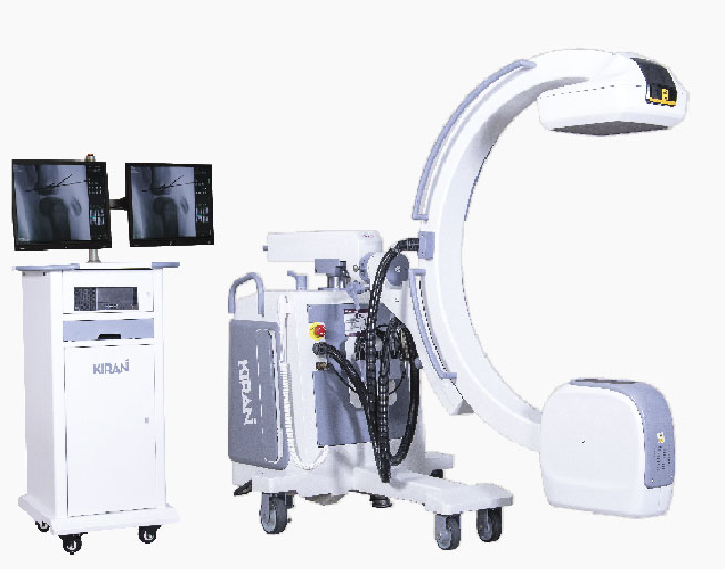
The C-Arm gets its name from the C shaped arm that holds an X-ray tube at one end and an Image Intensifier or a Flat Panel Detector at the other end. The Arm can be moved horizontally, vertically and can be rotated around the swivel axis to properly position the patient in the X-ray field and acquire the desired image. The console of C-Arm would generally house high voltage power electronics needed for the X-ray tube, control electronics for managing the C-Arm movement and embedded computer systems for image acquisition and processing.
C-Arm technology has evolved continuously since its introduction in 1955; and most recent technology trend is migration from Image Intensifier based Analogue technology to flat panel detector based digital technology.
Trivitron Healthcare is at the forefront of innovation in medical imaging using digital technology, having launched digital radiography, digital mammography and now being the leader in digital C-Arms.
In Analogue Image Intensifier C-Arms; the X-ray beam after penetrating the patient’s body hits the Input Phosphor end of the image intensifier; the input phosphor converts the X-ray to light photons which passes through a vacuum tube with an arrangement of Photo Cathode, Electrostatic Focussing Lens, PhotoAnode finally reaching the Output Phosphor end of the Image Intensifier and forms a visible image of the X-rayed body parts. This image is then captured by a CCD camera and gets transmitted to the display monitors.
In case of Analogue C-Arms, image conversion happens in two steps; Step 1: X-ray to visible light image conversion by the image intensifier; Step 2: Capture of visible light image by CCD camera and further processing using analogue means. Due to the curved surface of the Image Intensifier tube the accuracy of the image is diminished near the edges leading to distortion. Furthermore due to multiple steps and electron optics involved in the imaging chain; the field of vision is reduced with every step of magnification.
Flat Panel Digital Technology directly converts the X-Ray to an electrical charge which gets digitized in the detectors readout matrix. An Image Processing software converts the digital input from the detector to a digital image with a plethora of image processing options leading to high contrast and high resolution images that help visualize very minute anatomical structures. Digital C-Arms provide distortion free accurate images edge to edge of the entire viewing field.
Trivitron Healthcare offers a wide range of Digital C-Arm options; the Infinity series with 3.5 KW stationary anode and the Elite series with 5 KW rotating anode X-ray Monoblocs. Trivitron Healthcare C-Arms feature advanced software with Digital Subtraction Angiography, Roadmapping features along with dual panel or wide screen single panel display options.


